Crispin Weinberg | 3D printed patient-specific models for surgeons


Who is Crispin Weinberg?
Crispin Weinberg is an anatomical engineer & entrepreneur, and President of Biomedical Modeling Inc. , which produces 3D printed patient-specific models for surgeons and providing anatomical engineering services for medical device designers. Previously he served as Chief Scientific Officer of Angio-Oncology Sciences, Inc., co-founder of Organogenesis Inc., and Research Fellow at MIT. PhD (Neurobiology), Harvard University SM (Physics), SB (Mathematics), the University of Chicago.
In this episode we discuss the process on how to convert scan data to 3D CAD files, the benefits of proving surgeons with patient-specific models prior to surgery, and Crispin’s biggest success stories.
Biomedical Modeling Aids Guatemalan Conjoined Twins
EXPAND TO VIEW EPISODE TRANSCRIPTION
SUMMARY KEYWORDS
models, people, mesh, surgeon, scan, anatomical, solidworks, engineer, cad models, patient, podcast, area, ct scan, exported, file, software, devices, cad, called, density
SPEAKERS
Presenter, Crispin Weinberg, Aaron Moncur, Rafael Testai
Presenter 00:00
Hi everyone. We’ve set up this being an engineer podcast as an industry knowledge repository, if you will, we hope it’ll be a tool where engineers can learn about and connect with other companies, technologies, people, resources and opportunities. So make some connections and enjoy the show. The greatest moment of publicity, our 15 minutes of fame, so to speak, was the chief surgeon was interviewed on Good Morning America, and he came in he had our models in his lap.
Rafael Testai 00:45
Hello, everyone, welcome to the being an engineer podcast. We are co host Rafael Testai. Today we have another very special guests. Crispin Weinberg, who is an anatomical engineer and entrepreneur, the president of biomedical modeling, Inc, which produces 3d printed patient specific models for surgeons, and providing anatomical engineering services for medical device designers. He was previously a Chief Scientific Officer of angio oncology sciences, Inc, co founder of Organogenesis Inc. Organogenesis. I really appreciate the correction. Thank you. Yes. And research fellow at MIT, Ph. D. neurobiology, Harvard University physics, and SB mathematics from the University of Chicago. So Crispin, welcome to the show.
Crispin Weinberg 01:41
Well, thank you, Rafael. I’m delighted to be here. I think it’s a fascinating podcast, and I look forward to an enjoyable conversation with you.
Rafael Testai 01:50
Appreciate it. Is there anything else that I may have actually like when people correct me so I can improve? Anything else? I’m gonna have mispronounced in introduction?
Crispin Weinberg 01:59
I’m not sure you may have stumbled a little bit over anatomical engineer the first time. So I’d like to make that clear that its anatomical
Rafael Testai 02:08
list. Let’s start there. Because you are the first anatomical engineer that we have on the podcast. Could you define for us? What is an anatomical engineer?
Crispin Weinberg 02:18
Oh, great. Well, probably the first because I actually coined the term to try to describe what I do concisely. So an anatomical engineer, works primarily with medical imaging data and producing 3d models of it. We produce both physical models using 3d printing technologies, and virtual models of which can be a variety of formats from mesh models, which are more for visualization, to CAD models that you can use for simulation. And even things like CFD analysis.
Rafael Testai 03:01
So So I saw your website, I found you by looking up on Google 3d CAD biology and 3d CAD organs. And that’s how I came across your company biomedical modeling, Inc. So tell us about biomedical modeling, Inc. In very simple terms. What are the services you provide? And who your customers?
Crispin Weinberg 03:23
Oh, great. Yeah, so we provide several different types of services. First, and the way we started was to take medical imaging data, primarily CT scans or CAT scans, and produce 3d printed models for surgical planning prefabricating, prosthetics and training. You might wonder a little bit why do you need to do this? Surgeons often have a great deal of experience looking at medical imaging data. And but still, the way I put it is, we that is people have spent millions of years evolving to look at three dimensional objects and understand what they’re looking at. We’ve only spent the last 500 years evolving to look at two dimensional representations of three dimensional objects and understand them. So we’re not very good at it yet. Give us another number of 1000s or hundreds of 1000s of years and we won’t need the models. So that’s one aspect of models for surgeons. The second aspect is CAD models, primarily for medical device designers. Now probably most of our audience is familiar with CAD software and how it works, incorrect. Geometrical objects, circles and lines and ellipses of organic three dimensional shapes do not fit very naturally into CAD. If you try to import a mesh file into your CAD program. It wants to treat every single polygon triangle or quadrilateral depending on your mesh as a single face. And so you will immediately have this structure which has hundreds of 1000s or millions of faces. And it’s totally unworkable. So how do you get cat organic shapes into CAD. And basically, we go through that process, taking the mesh file, putting NURBS curves on it, exporting it as a STEP file and importing it into a CAD program, often with lots of back and forth to correct problems, because it’s easy to get self intersecting faces or other sorts of abnormalities. So you need to do that. And you also need to do that taking to count a little bit of what the final uses. So what you do for somebody doing a simulation might be very different than what you do for somebody doing a CFD study where they’re interested in the flow in one direction. So it’s not just a simple press a button, but it’s there’s a bit of a bit of art and experience to it as well. And so these are very useful for medical device designers who want to test their device on anatomical structures on human anatomy before they put it into people.
Rafael Testai 06:34
Let’s talk about the process of editing the mesh file for a little bit, because myself and a lot of my our audience here, we’re very familiar with CAD and a lot of them with SolidWorks. So once you take the CT scan, what will be the next step? How do you work the file?
Crispin Weinberg 06:51
Okay, well, again, the CT scan, just to refresh people’s memory is essentially a three dimensional X ray. And it’s usually exported as a series of images that are like slices, very much like you would slice a file for 3d printing. And what you want to do on that is analyze the data to put it together as a three dimensional model. So primarily look at the density of the CT scan, the density of the CT scan, because x rays reflects the density, the atomic numbers actually of the tissue that you’re looking at. So bone will appear up here, white, because it’s got a very high X ray density calcium as a high atomic number, and air appears black, because it’s got a very low density, most soft tissues appear various shades of grey. So we make essentially masks very much like you would do in Photoshop, based on density, and stack them together to make a three dimensional object, then editing involves cleaning that up, because if you try to pick all the regions of a certain density, if you’re looking at a very clear object, like a bone compared to soft tissue, it will probably do it fairly well. But when you start getting into more subtle objects in the soft tissue range, or some of the thinner bones, you end up getting places where you have to sort of manually go through and remove parts you don’t want connected or fill in little holes. Also, if you have metal objects, of course, they have very high atomic numbers. So they’ll produce artifacts. And you’ll see that as streaking and black areas. And so you’ll have to edit around those. So there’s a fair amount of understanding what you’re looking at in the CT scan, and making sure that your masks reflect that. And then you grow that into a three dimensional structure into a mesh. Or you can export it as a mesh that is actually in the medical imaging processing program. It’s actually a point cloud. And so but you can export that as a mesh by picking out that density. Usually that mesh is still very, has has problems that you need to fix. So the mesh represents the surface as a series typically of triangles as an STL file. STL originally stood for standard tessellation language and then sort of got pushed into being called stereo lithography language. So there’s a kind of ambiguity about what it stands for, but it means that you’re representing the source This as this large number of triangles, and a human skull, for example, might have 500,000 to a million triangles when you’ve exported it. So it has a lot, if you’ve just exported, imported those into SolidWorks, a lot of faces. So it’s a fairly complicated structure,
Rafael Testai 10:19
May I you question?
Crispin Weinberg 10:20
Sure, absolutely.
Rafael Testai 10:22
Once the MRI or CT scan is taken, you have data, the slices, and then you would the term be curate the data? whats the term?
Crispin Weinberg 10:32
the term that’s actually used the segmentation, we’re dividing it into the segments of different densities. So that’s officially what the, what the term is for, for describing that process of someone isolating three dimensional structures,
Rafael Testai 10:47
the segmentation, and What software do you do it?
Crispin Weinberg 10:52
we do it in a software called mimics, which is made by a company materialize in Belgium. That is pretty much the gold standard for segmentation software. There are others around. But most Li and they’ll work fine. But they don’t have the same suite of editing tools that allow you to your art, take care of artifacts and edit out artifacts effectively. And trace long skinny things like nerves. So that essentially, is a tool for drawing essentially a cylindrical spot spline along a path, if you can trace where a nerve runs
Rafael Testai 11:40
the social animal, a very great, very good explanation Sorry to cut you off. I’m just like every little by little, I’m trying to digest what you’re saying. It’s very technical, but you’re doing a wonderful job explaining it,
Crispin Weinberg 11:52
it is fairly technical, it’s also fairly visual. So talking about it on a podcast, that’s a little bit of a challenge, in a way. So like, I am enjoying trying to figure out how to articulate something. So when typically I explain it to people, I actually show them on the screen, well, here’s this area of density, and that’s a bone and this is a tooth, and this is something else that goes through a scan, and people get it very quickly. But an interesting point, if I can make a digression, okay, one of the one of the values of these models, to surgeons, and is also explaining to patients what they’re going to go through and what’s going to happen. A very interesting study was done early on, when the surgeon explained what the procedure was just verbally to a patient. They had this sort of modest understanding of what was going on, when they were shown pictures of the scan. And the surgeon said, Here’s your heart that when we’re going to do this, for example, their understanding actually went down because there’s this, they were just so mystified by what they were looking at, on the scan, that made their understanding go down. But when the surgeon used a 3d printed model to explain what was going on, their understanding went way up. And they were much happier about the procedure, and much more likely to do whatever follow up as necessary to improve their recovery because they had a much better understanding of what was going on. So they’re very effective tools for this sort of patient education as well.
Rafael Testai 13:38
Right on? Well, I want to go back first want to get down the process before we get into all the benefits. But unquestionably having a patient look at what’s going to happen or the biology of what the surgery is going to be like, I think it’s amazing and could provide a better understanding. But going back to the step of segmentation, using the Mimics software, once you finish that you import a mesh file into SolidWorks. They get there right
Crispin Weinberg 14:06
now then we go through, well, you could put it bring it directly try to bring it directly in but when you tried to do that, if if it has too many triangles, SolidWorks just crashes. The number of triangles you can import actually directly has been steadily going up. When we started doing this sort of work in with SolidWorks. I think we started working with it in like 2003 They couldn’t import very many mesh triangles at all. So numbers steadily going up but it still doesn’t really treat it that way. So what we do is we use a another process, essentially a reverse engineering software. We use Geomagic studio, but there were quite a variety of the them around. And basically, you take that you clean up the mesh, various tools are so smooth that so that the little irregularities don’t appear. The, if you actually export a very detailed mesh, you can see sort of stair steps for the individual slices of the CT scan. Obviously, the skull doesn’t, or whatever structure you’re looking at, doesn’t really have those stair steps. So you can smooth those out. And you can fill little holes, if you don’t need all that level of detail, or if they’re doing the artifact and sort of smoothed out the mesh. So it’s clean, you eliminate any places where there are self intersections, which may look fine if you’re looking at it just as an object on a screen. But if you try to 3d print it or do CAD analysis on it, you can’t do it, so you have to clean it up a bit. And that’s done in reverse engineering software. Then if we’re going to 3d printed, we can simply save it as an STL file and send that to a printer. If we’re going to do CAD on it, we then tried to simplify it by approximating the surface with curves that are laid on the surface called NURBS curves or non uniform B splines. If you’ve used Rhino, for example, you’re probably familiar with NURBS curves. But they essentially put like lines of latitude and longitude on the surface to approximate the shape of the surface. There’s definitely some loss of detail when you do that. And one of the things that we have to work on very closely with our customers is what level of detail they need preserved in a CAD model. Those NURBS curves can then be exported as a STEP file, and the STEP file can be imported into SolidWorks. Sometimes it comes in very cleanly, sometimes it comes in with lots of errors. And if you run import diagnostics in SolidWorks, you get along hideous less list of problems. And we sort of go back and forth between the reverse engineering software, and Solidworks. fixing the problems, usually if they’re relatively small until you can fix them in SolidWorks. But if you really have significant problems with the mesh structure, it’s generally easier to repair them as a mesh, and then import them into SolidWorks. I should also mention that when you’ve imported such a file into SolidWorks, you end up with something that doesn’t have a history tree, it’s just a solid, or what we sometimes call a dumb solid, because it has no history you cannot. And there are no sketches. You can’t edit it in any of consistently, you can push and pull on it, you can do combine it with other things. For example, if you want to make a model of an artery and put a hose BB on the end, so you can plug it into a model circulatory system, you can do that combination in SolidWorks and produce a file that you can then print that you can work with.
Rafael Testai 18:44
Very well explained. I think you mentioned that visuals are needed to well not needed but usually have visuals handy to explain this. But for everyone that’s listening to this driving. That was excellent. Thank you. My next question is going to be about something that you touched upon the level of detail of these files. What determines, of course, the customer will ask you, but in which cases do you need more detail than others?
Crispin Weinberg 19:11
It really depends on what a customer wants to do with it and is concerned about. So if you’re making a model that’s going to be used for designing dental work, for example, for orthognathic surgery, you need a fairly high level of detail in the teeth and lower levels in other structures. So you can actually make some regions very well detailed and others less, less highly detailed. If you’re interested in a blood vessels, you may be interested in branches which are larger than a certain diameter say larger than one millimeter diameter. I’ll so we could smooth out the various small branches, most of which we probably wouldn’t even see on the scan. But if there are any that we do see, we would be able to smooth those out. And then include all only the larger ones. So there’s really a lot of back and forth with the with the customers. And I think often I would say the most important criterion for the success of a project is a lot of communication with with a customer or client to make sure that we’re meeting their needs. Another, of course, restriction on the level of detail is the original data. The typical slices in a CT scan are anywhere from my half millimeter to five millimeters apart, depending on the scan, what a large area it covers. So we cannot improve on the resolution of the original scans. What are the difficulties with MRIs as opposed to CTS is that MRIs tend to have poor spatial resolution, they do have the advantage of much better soft tissue contrast. So if you’re interested in, for example, looking at the difference between fat and muscle, you would almost always want to use an MRI scan even though the spatial resolution is not as good
Rafael Testai 21:39
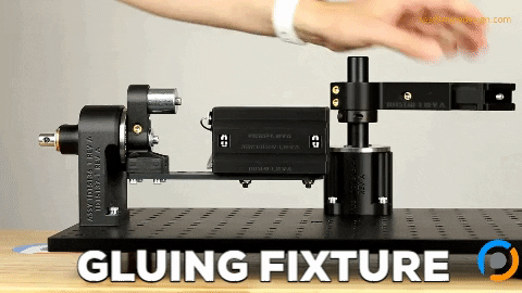
ski. I want to take a real quick break to mention to our listeners that this podcast is sponsored by pipeline design an engineer. And if you visit teen pipeline that US is where you can learn more about how we help medical device and other products engineering or manufacturing teams develop turnkey equipment, custom fixtures and automation machines to correct arise, inspect, assemble, manufacture and perform verification testing on your devices. Also a friendly reminder to our listeners that we really appreciate your podcast reviews. We’re trying to get to 105 Star podcast reviews on the App Store. It really helps other people find the podcast, I’m here with my guest, Crispin Weinberg. And my following question is going to be you mentioned several softwares are needed to edit the scan file before it’s delivered to the customer. So my question is, can one person is one person usually able to do all this all the all the editing? Or do you need multiple people on your team with different software skill sets to do this?
Crispin Weinberg 22:47
One person with a sufficient background and training can certainly do it. But often people will like to specialize in one area or another. So one of the ways I like to describe it as the what I do is probably about 50% engineering and 40% medicine and 10% art. The exact proportions may vary a little bit from one project to another. I certainly do. But there’s a there’s a mixture of skills. And one of the things that I’ve often done is, when looking for people, when I look at the resumes, I’m usually looking at biomedical engineers, but if they say anything about, you know, doing art projects of any sort, or ceramics, or by you know, particularly three dimensional art type things, I tend to be look very favorably on those. So that’s a very important criterion for trying to find people who can do this, it involves a very good sense of 3d vision, 3d visualization, which many, which most engineers have, of course, but also sort of artistic sense of trying to make it because you can make a three dimensional model. And if you just exported it from the medical imaging process, processing software, and built it directly is that it would probably look kind of ratty or ugly. But with a little bit of editing, you’re not actually changing the data, but you’re making it so it comes out looking much nicer. And one of the ways I describe what we do we make things that are both beautiful and terrifying at the same time. My favorite example of food and if you look at our website of the bio model.com There’s a picture of a large tumor in a jaw that we’ve colored red with a bone transparent. If you hold that up to the light, it’s beautiful. It glows like a Ruby. But at the same time, imagining this fist sized lump growing on somebody’s jaw is really terrifying and frightening and horrible. So it’s it’s a fascinating combination of things, which are both beautiful and terrifying.
Rafael Testai 25:28
So it sounds like you’ve been doing this for 20 years. Is that right? Bomb? Biomedical modeling increase. All right,
Crispin Weinberg 25:34
that’s right. Yeah, but just about 20 years.
Rafael Testai 25:37
Congratulations.
Crispin Weinberg 25:38
Well, thank you, as well.
Rafael Testai 25:41
I wanted to ask you, are you You said we, so it doesn’t sound like you’re a one man team. How big is your team?
Crispin Weinberg 25:48
Well, that’s actually pretty small. Really, right now, it’s just me and one other biomedical engineer and a part time bookkeeper. So it’s gotten pretty small. At times, it’s been up to like four or five people. But it’s not a huge business. Partly because I like to keep my hands on a lot of it. And a lot of it is this steady communication with the customers.
Rafael Testai 26:17
I see. Well, I want to ask you some questions about business just so we can get if anything’s off limits, just please let me know. But I want to get an idea of like, what’s your best seller in terms of what’s the CAD file that people tend to ask for the most often?
Crispin Weinberg 26:37
Oh, most people in terms of CAD files are probably looking at parts of the skeleton that is used, I think a lot in simulating how bodies function and respond to various accidents and devices and repairs, it’s also probably makes sense because the skeleton is rigid or unyielding and making physical models of it is pretty a good representation. So that’s probably the area where we’ve made the most CAD files. The second most is cardiovascular, which is of course, the complete opposite, it’s very soft and moving. But the heart, of course, is very complicated. And doing minimally invasive surgery, where people are feeding catheters up to the leg and into the heart and delaying procedures is an area that requires a lot of training and simulation to make sure your device can get around all those curves of those blood vessels and, and do its job. And so we’ve made quite a few CAD models for the heart, and particularly for minimally invasive mitral valve surgery.
Rafael Testai 28:10
Perfect. What’s what an what excites you the most about working in biomedical modeling Inc,
Crispin Weinberg 28:17
I think what really excites me is being able to use these engineering skills to make things that help people, it’s really wonderful to feel then part of making many of the devices which help improve medical care, and, and make it more more human I, one of the other areas that it’s can be very useful for, as is what’s starting to be called more personalized medicine, where you can actually customize devices for the individual patient. And as well as, for example, pharmaceutical treatments, because being customized for the individual patient. I think we’re we’re coming into an era where we recognize that different people have different metabolisms and different bodies and different anatomies. And they react differently to these things. So instead of going to this shelf and buying a hip implant, and saying, Well, I’m going to put in a one that’s either small, medium or large, you find one which fits the patient shape, and actually can make it there are a number of companies involved in doing things like that and starting to make products that are actually customized to the individual patient. And I think there’s we’re going to see much more of that in the next decade or two.
Rafael Testai 29:54
Fantastic if you were an 18 year old about to enroll in college is still you? What major would you choose? And why?
Crispin Weinberg 30:08
I probably would choose biomedical engineering right now, because I think it’s just fascinating. I think the way the body works is a marvelous, marvelous machine sets, let’s say, the way that different parts work together are just fantastic. And, and truly fascinating.
Rafael Testai 30:32
Has is impossible. And now we’re jumping subjects. But these questions are just popping up in my mind, is it possible to run F E, A analysis and some of the 3d scans you do?
Crispin Weinberg 30:44
Well, again, you don’t do FDA directly on the scan. But you can certainly do it on the on the CAD models that we’ve produced for from and we’ve certainly made a CAD models of for companies doing FDA type analysis.
Rafael Testai 31:04
Wonderful. Yeah, I misspoke on the graph is what I meant. All right. Well, is there something that I haven’t asked you that maybe I should have asked you that you want to share with the audience?
Crispin Weinberg 31:14
Well, I’d love to talk about some of the the special cases we’ve done. Probably the most famous case we have was actually just about 20 years ago now, which is hard to imagine. But it was the conjoined twins from Guatemala, who were joined at the back of the head. And they were going to be separated by a team at UCLA, they had to be separated, because they were joined at the back of the head. And eventually the brains would grow into each other and compress the two brains, and they would die from that probably somewhere around four or five, if they had managed to survive that long. So we got to make models for planning that surgery. And it was a very interesting experience. Because when we, we were weren’t initially involved in it. And the chief surgeon had had a resident who had worked with our models at a previous hospital where he had been, and he said to the surgeon, you should get these models. And a surgeon said, Man, why do I need that I can look at those scans. I know what I’m looking at. And fortunately, the resident persisted. He said, okay, okay. Let’s do it. But you know, we’re doing this case, pro bono, we don’t have any funds for it. And so they asked if we would donate our services and make piles. And we said, Well, yeah, there’s a very exciting, really challenging, so we agreed to donate models. And we made them, we made actually three models, which turned out to be very successful one model of each girl’s head, and then one model of the blood vessels between the brain. Well, the day that the surgeon received the models, he called us up and said, I saw something on the models that I hadn’t seen before in any of the images that I had looked at. And so first of all, it already showed that there are very powerful educational tools. And as I mentioned earlier, we’re used to looking at three dimensional objects and understanding what we’re looking at. When you’re looking at the two dimensional images, you tend to only look for the things you think you know, you’re looking for. And you don’t really notice the things that you’re not looking for, like the proverbial gorilla in the room sort of thing that you don’t look at those things because you’re not looking for them. When you look at that three dimensional objects, you see, oh, this part is twisted than their spines were twisted in a way that he did not know. That probably won’t make any difference to the operation. But it’s good to know, just in case it does. The next thing that we learned, well, the greatest moment of publicity are our 15 minutes of fame, so to speak, as the chief surgeon was interviewed on Good Morning America, about this procedure before it took place any payment, he had our models in his lap, and he picked them up with the two heads joined together. We put little alignment pins in there, and he told them apart. So this is what we’re going to do. And this despite the Vinay describe, of course, the models haven’t been made by our company biomedical modeling in Boston. And so this was incredible publicity for us, of course, and very excited. Probably also, the first time many people ever saw or heard of 3d printing, because at that time that was primarily something that was used by aerospace engineers and automotive engineers. And outside those areas, it was not known very widely. So it was also, I think, an important moment and publicity for our industry as well. Now, the whole industry of, of additive fabrication, then, the next great moment was when the operation took place, it was pretty big news story. And after the operation, which took, I believe it was 28 hours. Not everybody being there all the time, obviously. And we’ll get to that in a second. Then the all the big news media and so on, we’re trying to call a surgeon and interview him about Halloween. And he called us to thank us for the models, because he knew that he would probably still be in the operating room because it would have taken longer if they hadn’t had the models for planning. And the result might not have been as successful, the result was quite successful. Both girls survived, one with some neurological defects and one quite normal is remarkably good, because they actually shared blood vessels in the brain. So. So getting back to one of the other things I had mentioned, it was very fortunate, we sent the three models, We’d only been asked to make models of bone, the skulls. But it turned out the operation, in addition to having the chief plastic surgeon also had a chief neurosurgeon, who was going to do the dissection of the blood vessels. And so he had this model of the blood vessels, which helped him and particularly later, we’ve got video footage of the operation. And we saw that the chief neurosurgeon and the chief plastic surgeon, going off, getting together looking at the models and consulting and talking about what they were going to do, and then going back to the patient’s another use of the models had been for choreographing the operation, as I mentioned, there, the operation went on for a long time over the course of the operation are probably about 50 Different people at one time or another. They were also starting with one patient and ending up with two patients. So they essentially had to hold a dress rehearsal of who was going to be where, when, and the models were very useful as sort of serving as a surrogate that they could put have together and then pull apart for that. Another use, which was totally unanticipated was that the anesthesiologist use them who anesthetize somebody, you have to put a tube, a nasal gastric tube down their throat, drop the nose and down into the throat, to make sure they don’t aspirate, they don’t drown in their own fluid secretions when they’re unconscious. And normally, you do that by tilting a person’s neck back, so that you sort of straightened out the throat. And it’s much easier to insert a tube. Of course, with the girls whose were joined at the back of the head, it couldn’t bend their heads back in that way. And so she had to use the models to figure out how she was going to insert the nasal gastric tube, not something anybody had anticipated, as a problem, but it turned out that was solving that problem or helped solve that problem in advance. So anyhow, that’s a fairly long story about I could talk on and on about it. But it was an extremely exciting and gratifying thing to be involved in this procedure. And something that was very successful. And we got pictures from their 10th birthday party, which was really delightful. So we were very happy to be part of that.
Rafael Testai 39:20
Fantastic. Well, we’re gonna have links in the show notes for the articles that you reference your your 15 minutes of fame, which I thought was pretty funny. And yeah, hopefully we can drive some traffic to your website to make people aware of this wonderful resource. Any last words of encouragement for any engineers, maybe still in college?
Crispin Weinberg 39:43
I would encourage people to learn as much as you can about different fields. You never know how they’re going to fit together. You know, I would describe my as I stated when I describe myself now as an anatomical Engineer. Your that didn’t exist at all when I was in college, but by learning about physics and biology and mechanics, was in a position that I could come to, to do something and help develop a new field that moves things forward. So I encourage people to learn about many different things. Because many of you will be working in careers that don’t exist now, but will in the future, and it’s very exciting to be part of that, building the future for people. So thank you, Rafael. I think that’s a great.
Rafael Testai 40:40
Yeah, thank you so much for being on the podcast. I appreciate it.
Aaron Moncur 40:47
I’m Aaron Moncur, founder of pipeline design and engineering. If you liked what you heard today, please share the episode. To learn how your team can leverage our team’s expertise developing turnkey equipment, custom fixtures and automated machines and with product design, visit us at Team pipeline.us. Thanks for listening
We hope you enjoyed this episode of the Being an Engineer Podcast.
Help us rank as the #1 engineering podcast on Apple and Spotify by leaving a review for us.
You can find us under the category: mechanical engineering podcast on Apple Podcasts.
Being an Engineer podcast is a go-to resource and podcast for engineering students on Spotify, too.
Aaron Moncur and Rafael Testai love hearing from their listeners, so feel free to email us, connect on Facebook, Twitter, Instagram, and subscribe on Apple Podcast and Spotify!
About Being An Engineer
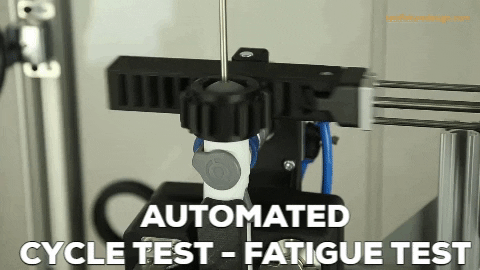
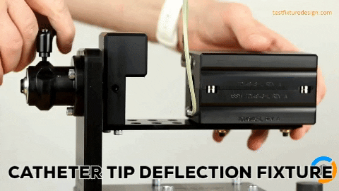
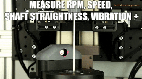
The Being An Engineer podcast is brought to you by Pipeline Design & Engineering. Pipeline partners with medical & other device engineering teams who need turnkey equipment such as cycle test machines, custom test fixtures, automation equipment, assembly jigs, inspection stations and more. You can find us on the web at www.teampipeline.us.
You’ve read this far! Therefore, it’s time to turn your headphones up and listen now to this episode to learn all these. Don’t forget to tell your friends who might like this too!